Need for frequent BP monitoring in unstable patients
Need for frequent blood gas measurements in patients on a ventilator
If venous access is limited and multiple lab draws are needed
Third choice of arterial line sites behind radial and brachial in light of increased infection risk
CONTRAINDICATIONS
Severe coagulopathy; platelets<50k; INR>1.5-1.6 (relative contraindication)
Evidence of impaired perfusion with significantly decreased pulse
Infection/inflammation over site
Presence of arterial graft at site to be used (Fem-Fem bypass, etc)
RISKS
Thrombosis/Bleeding
Infection
CHOICE OF ARTERIAL LINE SITE
The radial artery is most often used; advantages include ease of placement, relative accuracy, presence of collateral flow. Note that if a short catheter is used in the radial position, BP may be underestimated on high dose vasopressors in septic patients. Vasopressor effect on the longer catheter normally used is unknown.
Brachial artery catheters are another option, and should be located just above the elbow crease. The technique is the same as for radial placement.
Dorsalis pedis artery catheters may also be placed as an alternative; a short catheter may need to be used in this situation.
The femoral artery is an option that is often employed when radial catheters cannot be placed. It is a relatively easy artery to cannulate, and may be more accurate in sepsis when high dose pressors are used. Disadvantages include an increased risk of infection and it may be problematic when a patient is awake and moving the legs.
TECHNIQUE
Ensure there is an adequate pulse in the femoral artery prior to attempting the procedure.
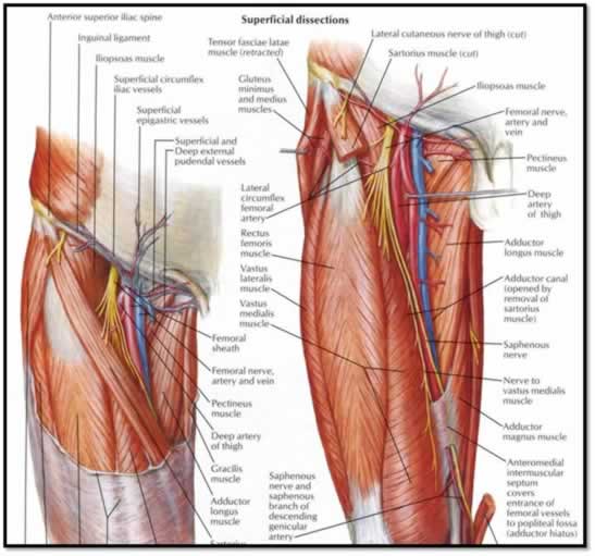
Review Anatomy, from lateral to medial "NAVL", "venous penis"
Prep an area over the femoral artery about 15-20 cm inferior to the inguinal ligament, and cover with the drape provided.
Anesthetizing the area over the artery with lidocaine helps in comfort and may reduce arterial spasm. Too large a wheal can obscure the artery, so keep it small.
Make sure to insert the line 1-2cm below the inguinal ligament to assure you don't enter the retroperitoneal space.
While palpating the artery with your non-dominant hand, use the large finder needle to advance through the skin at a 30 degree angle.
When the artery is entered, a pulsatile flow of blood will be seen.
Once in the artery, advance the guide wire through the needle, and remove the needle, always making sure to be holding on to the guide wire. If the guide wire will not thread, remove it and the needle, and try a different spot.
For a femoral arterial line, always use the long (12cm) catheter.
Place the 12 cm catheter over the guide wire, and advance until the hub is up to the skin.
Remove the guide wire, and connect the catheter to a stopcock for measuring. See if an arterial tracing is obtained.
Suture the sides of the catheter to the skin to ensure it doesn't fall out.